How is a pleural effusion diagnosed?
Diagnosis aims to confirm that fluid is present, estimate its volume and identify the underlying cause. The healthcare team will consider your symptoms, medical history and examination findings before selecting suitable tests.
Medical history and physical examination
Your doctor will ask about breathlessness, coughing, chest pain, fatigue, previous illnesses and current treatment. During the examination, the chest may be listened to and tapped. Reduced breath sounds or a dull sound on percussion can indicate fluid.

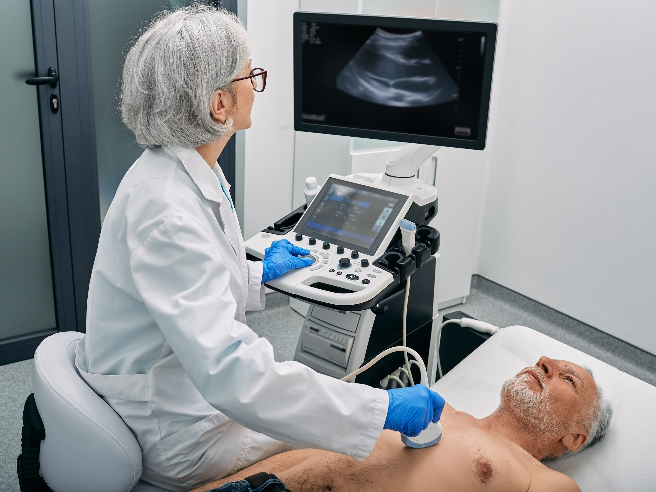
Ultrasound
Chest ultrasound can detect even relatively small amounts of pleural fluid and helps determine its location and depth. It is also commonly used to guide thoracentesis or catheter placement, helping the clinician select a safe access point.
Chest X-ray
A chest X-ray can show a larger pleural effusion and how it affects the lung. Depending on the amount and distribution of fluid, typical changes may be visible at the lower edge of the lung or across a larger part of the chest.
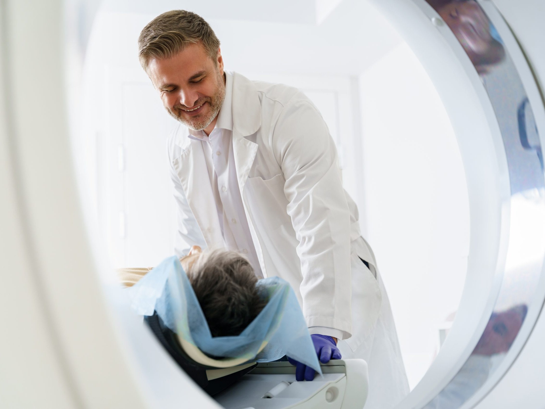
Computed tomography and magnetic resonance imaging
Computed tomography (CT) provides detailed images of the lungs, pleura and surrounding structures. It can help identify tumours, inflammation, thickening of the pleura or other possible causes. Magnetic resonance imaging (MRI) is used less often but may be helpful in selected situations.
Thoracentesis and pleural-fluid analysis
During thoracentesis, a needle or small catheter is inserted into the pleural space under sterile conditions and fluid is removed. This can relieve symptoms and provide a sample for laboratory analysis.
The fluid may be examined for protein and other chemical markers, inflammatory cells, bacteria and tumour cells. These results help distinguish between different causes, such as heart failure, infection or malignant disease.
Blood tests and further investigations
Blood tests can provide information about inflammation, organ function, protein levels and other possible causes. Depending on the findings, additional investigations may be needed, for example cardiac assessment, microbiological tests, bronchoscopy or a tissue sample from the pleura.

Conclusion
A pleural effusion has many possible causes, so a structured diagnostic assessment is essential. Imaging confirms the fluid, while analysis of the pleural fluid and other tests help identify the underlying condition. Your healthcare team will explain which examinations are appropriate for you.
If you have questions, please contact us or speak with your healthcare team.